Otology
NECROTIZING OTITIS EXTERNA
1.NECROTIZING OTITIS EXTERNA ( Short note 2003)
A.k.a: Malignant Otitis Externa
DEFINITION:
Rare complication of Otitis Externa
Severe infection of temporal and adjacent bone
May lead to osteomyelitis of temporal bone and skull base
Commonly caused by Pseudomonas aeruginosa
Typically seen in immunocompromised and DM patients
COHEN DIAGNOSTIC CRITERIA:
| MAJOR:
|
MINOR:
|
| A- MicroAbscess | A – Age (elderly) |
| B- Bone scan positive | B – Bacterial culture (pseudomonas – 98%) |
| C- Cruciating pain | C – Cranial nerve palsy |
| D- Discharge | D – DM/ debilitating illness |
| E- Edema | E – ESR raised |
| G- Granulation tissue | |
| F- Failed medical therapy |
DIAGNOSIS:
Lab: ESR, swab c&s, HPE of granulation tissue, TWC, Glucose, Creatinine level
Imaging:
CT scan – to exclude temporal bone SCC. Findings: soft tissue enhancement in EAC +/- abscess
Technetium 99 – for diagnosis (absorbed by osteoclast and osteoblast)
Galium 67 – for prognosis (absorbed by macrophages)
TREATMENT:
Medical – IV abx (anti-pseudomonal): Ciprofloxacin 1/12, sugar control, pain control, ear toileting
Surgical – mastoidectomy + debridement of granulation tissue
Sudden Sensorineural Hearing Loss
- labyrinthitis
- autoimmune disease (eg: Cogan’s syndrome)
- Acoustic neuroma grows 1mm/year grow ,unilateral (bilateral in Type 2 Neurofibromatosis)
- Meningioma
- Vestibulotoxic: gentamicin(aminoglycoside), dietatic, aspirin, quinolone(antimalaria)
- Cochleotoxic: streptomycin, kanamycin,neomycin
- FBC – anemia, polycythemia, high twc
- ESR
- RP
- Fasting serum lipid
- FBS, HbA1c
- Autoimmune screening c3/4
- TFT -hypothyroid
- VDRL -syphilis
- PTA
- MRI- do later
- DM patient – monitor sugar as steroid started, vertigo, logistic —> treat in patient
- Others treat as outpatient
- inject dexamethasone through TM under LA (use EMLA) n lie down for 30 minutes- every week once for 3 weeks
- dose: 4 mg ( 1ml) each injection
- Age: Young <10 years old and >60 years old: bad
- Esr > 25: bad
- Vertigo: bad
- Comorbid: bad
- Severity: Severe to profound HL: bad, mild / moderate: good
- Bilateral: bad
- Tinnitus: good (protective)
Tympanic membrane retraction
Tos classification of pars flaccida: (Tos et al)
- Grade I: Simple / slight retraction (not adhered to malleus)
- Grade II: Adhered to neck of malleus (fundus seen)
- Grade III: Hidden retraction pocket (fundus not seen)
- Grade IV: Erosion of scutum (outer attic wall)
Sade classification of pars tensa: (Sade et al)
- Grade I : Mild TM retraction
- Grade II : TM retraction in contact with incus or stapes (tympanoinducoplexy)
- Grade III : TM in contact with promontory wall (not adhered)
- Grade IV : TM adhered to promontory (atelectatic)
- Grade V : Type III or IV with TM perforation
Cholesteatoma (Part 2)
Histopathology:
Macroscopic:
Cyst like structure in ME cleft that contains pale debris
Microscopic:
- Fully differentiated stratified keratinizing squamous epithelium (capsule/ matrix)
- A nuclear keratin squamous epithelium (core)
- Epidermal Langerhans cells
- FB granuloma/ FB Giant cells
- Polyps
- Granulation tissue
How does squamous epithelium in cholesteatoma differs from skin? [viva]
- Absence of skin appendages ( glands/ hair follicles)
Different types of operation for cholesteatoma:
Canal wall up procedure :
- Atticotomy
- Atticoantrostomy
- Cortical mastoidectomy
- Combined approach tympanoplasty
Canal wall down procedure :
- Modified radical mastoidectomy
- Radical mastoidectomy
Causes of persistent ear discharge post mastoidectomy:
- Infection
- Residual
- Recurrence
- High facial ridge
- Granulation tissue/ mastoid cavity not well epithelialized
Sites of residual cholesteatoma/ difficult areas of cholesteatoma:
- Facial recess
- Sinus tympani – area medial to facial nerve.
To overcome: also do anterior tympanotomy for better access.
- Sinodural angle
- Mastoid tip
- Zygomatic root
- Anterior epitympanum – due to head of malleus blocking the view (posterior tympanotomy access)
Difficulty/problem in Down syndrome:
- Atlantoaxial dislocation
- Heart disease
- Short neck, macroglossia – difficult in anaesthesia
- Narrow airway, reduced tone
Why do Down’s syndrome have persistent discharging ear?
- Unable to complain
- Small EAC
- ET dysfunction – patulous ET ( no angle)
- A/w cleft lip and palate
- Difficult to cooperate for ear toilet and examination
Cholesteatoma
Definition: (Abramson et al, 1977)
Points to mention: [viva]
- 3 dimensional epidermal and connective tissue structure
- Forming a sac
- Conforming the middle ear cleft ( middle ear, attic, mastoid)
- Capacity for progressive and independent growth involving the underlying bone and replacing the middle ear mucosa
- Tendency to recur

Classification:
- Congenital
- Acquired
- Primary (no previous h/o ear infection)
- Secondary (previous h/o ear infection)
Congenital cholesteatoma aka: epidermoid cyst
Definition:
Squamous epithelial cyst that can arise anywhere within the temporal bone
LEVENSON CRITERIA (5)
- White mass medial to TM
- Normal pars tensa and pars flaccida
- No history of otorrhea or perforation
- No history of previous ear surgery
- Prior bouts of OM not ground for exclusion

Theories for development of Cholesteatoma (6) [viva]
- Congenital cell rests ~ epidermoid formation in the anterior epitympanic area in the developing fetal middle ear
- Metaplasia of middle ear epithelium ~ squamous cell metaplasia in the ME epithelium in patients with cholesteatoma
- Papillary ingrowth through an intact TM at the Prusak space.
Why Prusak space? Answer: Poor ventilation in the area.
4. Invagination of epithelium through pre-existing retraction pocket or perforation
– most widely accepted theory
– obstruction of Eustachian tube causing impaired ME ventilation —> TM retracted and forming retraction pocket. Continuous desquamation and keratin accumulation leads to cholesteatoma formation.
5. Implantation theory
– iatrogenic: grommet insertion/ Tympanoplasty –> implantation of squamous cell into ME
6. Epithelial invasion theory
– invasion of skin from meatal wall through marginal or attic perforation
Management of attic cholesteatoma [viva]
– able to see fundus: conservative management / ear toileting
– not able to see fundus: do PTA and HRCT
PTA shows >40dB HL: possible ossicular chain involvement
HRCT : anatomical/ surgical landmark & disease extent
- Mastoid pneumatization – well pneumatization/ sclerotic
- Facial nerve dehiscence
- Ossicular disruption/ erosion
- Tegmen tympani/mastoidea breach
- Lateral SCC erosion

*** pictures are not mine. I googled them.
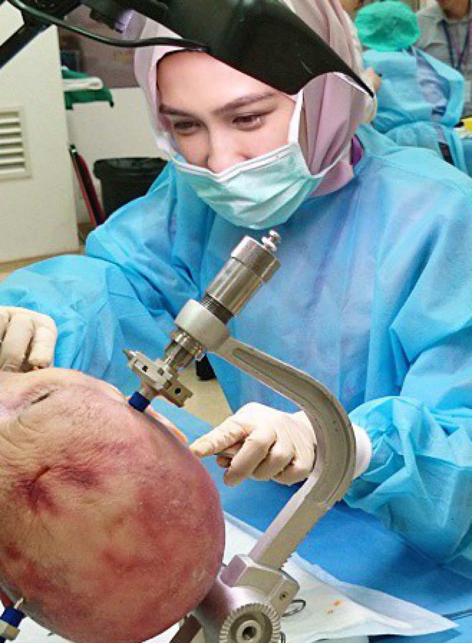
43rd UKM Temporal Bone Dissection Course 2015
On 22nd till 24th April 2015 I attended the 43rd UKM Temporal Bone DIssection Course 2015. This course is intended for otolaryngologists and residents/fellows in training to learn more about the temporal bone anatomy, related diseases and the management and also applied surgical techniques.
The venue of this course took place at the HUKM ORL Dept Level 9. The price was RM1000. That was quite a lot of money although luckily I managed to find sponsor.
Topics covered:
- Anatomy of the temporal bone
- Surgery for chronic ear disease
- Conductive hearing loss
- Surgery for otosclerosis
- Sensorineural hearing loss
- Cochlear implants
- Acoustic neuroma
- Surgery for acoustic neuroma
Procedures demonstrated and performed:
- Cortical mastoidectomy
- Endolymphatic sac decompression
- Facial nerve identification
- Stapes surgery
- Canal wall down mastoidectomy
- Cochlear implant
- Labyrinthectomy
- Translabyrinthine approach to the skull base
Overall I think this course was really beneficial for my learning. I was so caught up drilling the temporal bone that I almost always ended up didn’t realize how fast the time past. I wish we were given more time 🙂


